
Blog
Dental hygiene tips for healthy teeth & gums
What Is a Panoramic Dental X-Ray and Why Is It Done?
Why Dentists Sometimes Need A Bigger Picture
During many dental visits, the dentist begins with a quick look inside the mouth. The gums are checked. Each tooth is examined carefully. Dentists may take small X-rays as well. They help in many cases, yet they cannot always show what is happening deeper in the mouth.
Some dental issues develop deeper in the jawbone. Others appear around teeth that are still forming. At times, a wider view of the mouth is necessary. Dentists may then take a panoramic dental X-ray.
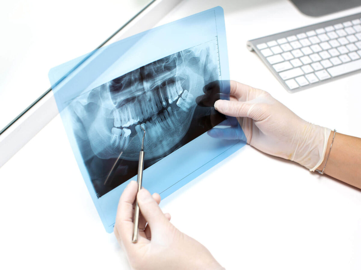
This image shows the entire mouth at once. The jaw joints, wisdom teeth, and surrounding bone can all appear in the same scan.
What A Panoramic Dental X-Ray Actually Shows
A panoramic dental X-ray works differently from the small X-rays dentists sometimes take. Those focus on a few teeth. This one shows the whole mouth at once. The machine moves around the head and records a curved image of the jaws and teeth.
The final image includes both rows of teeth. It also includes the jawbone. The sinuses above the upper teeth can appear as well. Jaw joints near the ears are often visible too. Seeing all of this together helps dentists understand how the mouth is structured.
Some changes in the mouth happen slowly. A broader scan can help bring those details into view.
Inside the Dental Panoramic X-Ray Process
A dental panoramic X-ray works a bit differently compared with the small X-rays dentists sometimes take. Those usually involve placing a sensor inside the mouth. This scan works another way. The machine moves gently around the patient’s head instead.
During the scan, patients usually remain upright. They place their chin on a small support. The head needs to stay still for a moment. Then the imaging arm slowly moves around the face as the scan begins. It records the images as it moves.
The entire process is quick. Often it takes less than a minute. Most patients find it comfortable since nothing has to be placed inside the mouth.
Why Dentists Use Panoramic X-Ray Teeth Imaging
Dentists rely on panoramic X-ray teeth images for several reasons. One of the most common uses involves evaluating wisdom teeth. These teeth often develop deep within the jaw before they become visible in the mouth.
The wider image helps dentists see where the wisdom teeth are positioned. It also shows the angle at which they are growing. Sometimes a wisdom tooth may be trapped under the gums. In other cases, it may begin to grow sideways. When that happens, the image gives dentists helpful clues for planning treatment.
Panoramic images can also reveal other concerns. Jaw fractures may appear in the scan. Bone irregularities can show up as well. Dentists may also notice cysts forming near the roots of certain teeth.
The American Dental Association explains that dental radiographs help reveal conditions that cannot be seen during a visual exam alone.
When Dentists Usually Recommend This Type Of X-Ray
A panoramic scan is not needed during every dental appointment. In many situations, dentists rely on a visual check and smaller X-rays. But occasionally the dentist needs a much wider view. That is when a panoramic dental X-ray may be suggested. Instead of showing only a few teeth, it captures the entire mouth in one image.
This type of scan is often taken before orthodontic treatment. Orthodontists spend time studying the image. It shows how the teeth are positioned. It can also reveal how the jaw is developing.
Sometimes, teeth that have not erupted yet appear in the scan.
The same scan may be used before implants. Dentists also use it when planning oral surgery. A full view of the jaw helps them prepare more accurately.
What The Experience Feels Like For Patients
Hearing the word “X-ray” can make some patients uneasy at first. Many imagine something uncomfortable. The panoramic scan, however, is usually quick and painless.
For the scan, the patient is asked to stay still briefly. The machine gently moves around the head as the image is recorded. The motion is gentle and quiet. Most people barely notice it happening. In fact, the entire scan usually finishes within a few seconds.
Because nothing needs to be placed inside the mouth, many patients say the dental panoramic X-ray feels easier than traditional bitewing X-rays.
Is A Panoramic Dental X-Ray Safe
Dental imaging today involves only a small amount of radiation. The levels are carefully controlled. Modern machines also include safety features that help limit exposure during the scan. For most patients, the amount is very low.
X-rays are usually taken only when dentists feel they are necessary. A routine exam does not always show everything. Some problems develop below the gums or deeper in the jawbone. Imaging helps dentists notice concerns that might otherwise remain unseen.
Dental imaging has been studied for a long time. The National Institute of Dental and Craniofacial Research has also discussed it in its research. Their research indicates that the imaging is considered safe when modern equipment and proper techniques are used.
What Dentists Look For In The Image
After the image appears on the computer screen, the dentist begins reviewing it carefully. Different areas of the mouth are reviewed one by one. The jawbone usually receives careful attention. Bone health matters because it plays a role in keeping teeth stable over time.
The dentist also checks how the teeth are spaced. Sometimes teeth grow too close together. At times, teeth remain beneath the gums instead of erupting normally. When dentists can see the full jaw in a single image, these situations become easier to identify.
Other structures often appear as well. The sinuses may be visible above the upper teeth. Jaw joints can also be seen in the image. At times, these areas may reveal conditions related to facial pain or bite problems.
How Panoramic Imaging Helps With Early Detection
Early detection is one reason dentists rely on panoramic X-ray teeth imaging. Many dental concerns begin quietly and develop over time. In the early stages, people often feel nothing at all. No pain. No clear symptoms.
The wider image makes a difference. It allows dentists to see areas that usually stay hidden during a routine exam. Subtle changes in the bone may appear. Cysts can sometimes show up as well. Occasionally, the scan even reveals unusual growths before they begin causing noticeable problems.
FAQs
Why do dentists take this type of scan?
The scan provides a wider picture of the mouth. Teeth, jawbone, and nearby areas can appear together in the same image.
How is this scan different from smaller X-rays?
Smaller X-rays focus on a few teeth. This scan shows the entire mouth and both jaws at once.
Does the scan cause discomfort?
Many patients say the process feels easy. The scan is quick, and nothing has to go inside the mouth.
How often is it usually taken?
Not at every visit. Dentists suggest it only when they need a broader view of the mouth or jaw.
Conclusion
Dentists rely on imaging more than ever today. A visual exam can show quite a bit, but it does not always reveal what is happening below the gums or inside the jaw. Some concerns remain hidden for a while.
That is where a panoramic dental X-ray becomes useful. The scan captures a broad image of the mouth and jaw in one view. With that wider perspective, dentists can identify potential problems and think through treatment options.
Sometimes a dentist recommends a panoramic dental X-ray simply to get a wider view. A routine exam does not always show everything. Certain problems develop deeper in the jaw or around teeth that are not easy to see.
The scan is quick. Most patients are surprised by how fast it is finished. Yet the image often provides helpful details that make diagnosis and treatment planning easier.

